


https://micro.magnet.fsu.edu/primer/techniques/fluorescence/gallery/cells/mrc5/mrc5cellslarge6.html
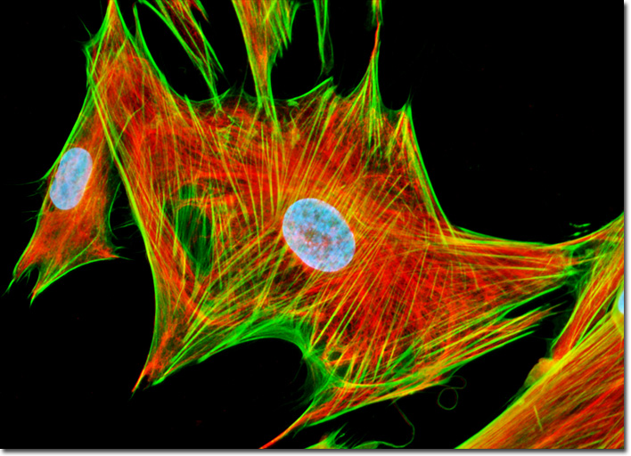
For this cell project I decided to choose a human fetal lung fibroblast cell from FSU’s fluorescence digital image gallery. The cell was taken from a 14 week old human fetus’ lung tissue. Fibroblast cells are the most common cells in connective tissues. These tissues are what connects and supports the organs in the body. Fibroblast cells play a major role in healing. In the cell image, the cell is dyed in three different colors; blue, red, and green. The cells pictured in image were treated with mouse anti-vimentin primary antibodies and goat anti-mouse secondary antibodies. These antibodies were dyed red, the nucleus is blue, and the Filamentous actin is green. Actin are involved in cell movement, signaling, and shape.
I chose the human fetal lung fibroblast cell because I have allergy induced asthma. There are times when my asthma is just a minor annoyance, however there are also times when it can be very severe. If I am not careful and unprepared for an asthma attack, this could result in a trip to the hospital. I see my asthma as a defect in my lungs and it affects my daily life. I thought it would be interesting to build a model of a cell that makes up my lungs.
Because fibroblast cells are in connective tissues, they attach to each other. In the image, you can see the cell at the center has three other cells connected to it by the filamentous actin, which is the outer, highlighted layer of the cell. For my cell form made of wire, I chose to construct the three layers present in the fluorescent image. In order to translate my cell into a 3 dimensional structure, I started building my cell from the inside out. I first started with the nucleus, using the 16 gauge wire as a base and wrapping it in 24 gauge wire. Next I built the antibodies, which I only used copper 24 gauge wire. I wanted the three layers to be distinct, therefore I chose to include copper wire. Finally, I made the outer actin. Because it needed the most support, I made a frame out of the thicker 16 gauge wire. In order to make it into a 3 dimensional form, I connected two pieces of bent wire diagonally on both sides of the frame. Finally, I attached the 24 gauge wire to the pointed ends of the cell frame in order to hold the inner layers in and to mimic the green actin in the image.
I think that I successfully translated the fluorescent image into a 3 dimensional structure. I was able to create all three layers of the cell shown in the cell image. Because of my research of the the fibroblast cell, construct the cell’s shape. Something I would change about my wire cell model is to make it so that it can stand upright instead of on the back of the cell. This would allow the viewer to clearly see the three different layers of the cell.
https://www.ncbi.nlm.nih.gov/pmc/articles/PMC3130349/
https://en.wikipedia.org/wiki/Fibroblast
https://micro.magnet.fsu.edu/primer/techniques/fluorescence/gallery/cells/mrc5/mrc5cellslarge6.html